Call us now :07971405282

 Send Inquiry
GEL DOCUMENTATION SYSTEM
MOQ : 1 Piece
GEL DOCUMENTATION SYSTEM Specification
- Shape
- Rectangular
- Glass Type
- UV-transmitting filter glass
- Accuracy
- High resolution imaging
- Temperature Resistance
- Ambient to 40C
- Features
- High sensitivity camera, UV protection window, easy sample placement, integrated image capture software
- Control Type
- Touch control panel
- Power Supply
- 230V AC, 50 Hz
- Display Type
- Digital/LCD display
- Usage
- Laboratory
- Type
- GEL DOCUMENTATION SYSTEM
- Dimension (L*W*H)
- 350 x 300 x 410 mm (approx)
- Product Type
- GEL DOCUMENTATION SYSTEM
- Equipment Type
- Laboratory Gel Documentation System
- Equipment Materials
- Aluminum enclosure with acrylic front door
- Power
- 230V AC, 50 Hz
- Voltage
- 230V
- Material
- Aluminum & Acrylic
- Application
- For documentation and analysis of DNA, RNA, and protein gels
- Warranty
- 1 Year
- Color
- White (as per image)
- Weight
- Approx. 12 kg
- Filter Options
- UV, amber, and optional blue light filters
- Imaging Area
- Approx. 20 x 20 cm
- Safety Features
- Auto UV shutoff, protective viewing window
- Light Source
- UV Transilluminator, white LED optional
- Software Compatibility
- Windows compatible software provided
- Connectivity
- USB interface, SD card slot
- Camera Resolution
- 5 to 12 Megapixels (depending on model)
- Image Output Format
- JPEG, TIFF, BMP
GEL DOCUMENTATION SYSTEM Trade Information
- Minimum Order Quantity
- 1 Piece
- Supply Ability
- 100 Pieces Per Month
- Delivery Time
- 7 Days
About GEL DOCUMENTATION SYSTEM
* Fast and easy-to-use basic gel documentation system for nucleic acid and protein gels
* Tray-based system for application driven image acquisition
* Small benchtop footprint
Image Acquisition
CCD camera-based system with four application-specific trays:UV tray for DNA and RNA gels and fluorescence stain imaging
White tray for Coomassie, copper, silver, and other EPI white light applications
Blue tray for low-intensity UV DNA/RNA detection (avoid "nicking" DNA)
Stain-free tray for in-gel protein detection
High-Sensitivity Imaging and Flexible Documentation
Engineered with a sensitive camera ranging from 5 to 12 megapixels and a spacious imaging area, this system enables precise capture and analysis of gels. Switchable filters and advanced lighting options cater to various staining and imaging needs, delivering high resolution images for DNA, RNA, or protein studies.
User-Friendly Operation and Safety Controls
A touch control panel, digital/LCD display, and Windows-compatible integrated software create an intuitive workflow from image capture to export (JPEG, TIFF, or BMP). Safety features such as automatic UV shutoff and a protective viewing window enhance operational security and convenience.
Durable Construction and Versatile Connectivity
Constructed from robust aluminum with an acrylic front door, the enclosure withstands routine laboratory conditions. The Gel Documentation System offers both USB and SD card connectivity, making data storage and transfer seamless. The equipment is designed for laboratories, research facilities, and educational institutions across India.
FAQs of GEL DOCUMENTATION SYSTEM:
Q: How does the auto UV shutoff feature protect users during operation?
A: The auto UV shutoff automatically deactivates the UV light source when the protective door is open or after a predetermined period, minimizing exposure and enhancing operator safety while handling gels.Q: What is the benefit of having multiple filter options such as UV, amber, and blue light in the system?
A: Multiple filter options allow compatibility with a range of nucleic acid and protein stains, such as EtBr, SYBR Safe, or Coomassie, ensuring clear visualization and documentation for different applications.Q: When is the system most effectively used in the laboratory?
A: The Gel Documentation System is most effective after electrophoresis, when rapid, accurate imaging and analysis of DNA, RNA, or protein gels are required for downstream applications like quantification, archiving, or publication.Q: Where can images captured by the system be stored or exported?
A: Captured images can be saved directly to an SD card or transferred to a computer via USB, accommodating flexible data management and integration with laboratory workflows.Q: What is the process for placing and imaging a gel with this system?
A: Open the acrylic front door, position the gel on the UV transilluminator, select the suitable filter and illumination, secure the door, and use the touch control panel to capture and view the high-resolution image on the digital/LCD display. Images can then be saved or exported as needed.Q: How does the integrated software enhance data analysis and workflow?
A: The Windows-compatible software streamlines image acquisition, analysis, and export, allowing users to annotate, quantify, and securely manage gel data efficiently without the need for third-party programs.Q: What usability features make this system suitable for laboratory environments in India?
A: The systems durable aluminum-acrylic construction, high-resolution output, easy sample placement, and compatibility with standard power (230V AC, 50Hz) ensure reliable performance in laboratory and educational settings across India.
Tell us about your requirement

Price:
Quantity
Select Unit
- 50
- 100
- 200
- 250
- 500
- 1000+
Additional detail
Mobile number
Email

More Products in Biotechnology Lab Category
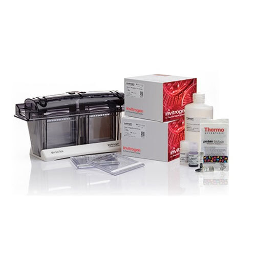
Electrophoresis Systems
Minimum Order Quantity : 1 Piece
Material : High Grade Polymer & Glass
Warranty : 1 Year
Type : Other, Electrophoresis Systems
Usage : Laboratory
Product Type : Electrophoresis Systems
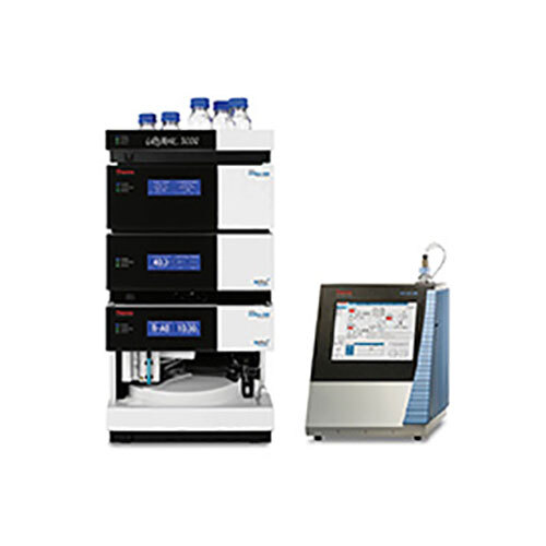
Chromatography System &Columns
Material : Stainless Steel, Glass
Warranty : 1 Year
Type : Other, Chromatography System &Columns
Usage : Laboratory
Product Type : Chromatography System &Columns
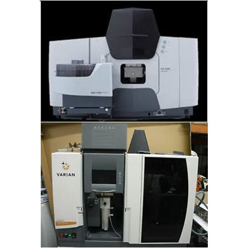
Atomic Absorption Spectrophotometer
Minimum Order Quantity : 1 Piece
Material : Mild Steel
Warranty : 1 Year
Type : Other, Atomic Absorption Spectrophotometer
Usage : Laboratory
Product Type : Atomic Absorption Spectrophotometer
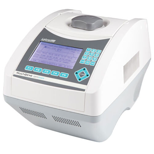
THERMAL CYCLER
Minimum Order Quantity : 1 Piece
Material : Plastic
Warranty : 1 Year
Type : Other, THERMAL CYCLER
Usage : Laboratory
Product Type : THERMAL CYCLER
Our Products
- Quality Control Lab
- Biotechnology Lab
- Life Science & Biochemistry
- Nanotech Lab
- Medical & Biomedical Lab
- Metal Analyzing
- Pharmacy Lab
- Industrial Chemicals
- Foundry Alloys & Metals
- Micronutrients For All Crops
- Glasswares
- Lab Furniture
- EDP Equipments
- LABORATORY CHEMICALS
- Mechanical engineering
- Civil engineering
- Electrical/Electronics engineering
- Electronics & Communication engineerings
- Textile Technology
- aerospace/aeronautical engineering
- Automobile engineering

160, 6th Street Extension, 100 feet road, Gandhipuram, Coimbatore - 641012, Tamil Nadu, India

Mr C. S. R. Cholan
(Chief Executive Officer)
Mobile :07971405282
 Send Inquiry
 Send SMS
 Call Me Free
Developed and Managed by Infocom Network Private Limited.
