Call us now :07971405282

 Send Inquiry
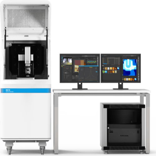
CONFOCAL MICROSCOPE
MOQ : 1 Piece
CONFOCAL MICROSCOPE Specification
- Temperature Resistance
- Up to 40C
- Accuracy
- 0.1 m
- Display Type
- High-definition touchscreen display
- Speed Range
- 500 fps acquisition speed
- Shape
- Rectangular
- Glass Type
- High precision optical glass
- Control Type
- Computer controlled (Software interface)
- Features
- Laser Scanning, Fluorescence Imaging, Z-Stacking, Automated Focus, Multi-Channel Imaging
- Power Supply
- AC power supply, 220V/50Hz
- Usage
- Laboratory
- Type
- CONFOCAL MICROSCOPE
- Dimension (L*W*H)
- Approx. 600mm x 950mm x 650mm
- Product Type
- CONFOCAL MICROSCOPE
- Equipment Type
- CONFOCAL MICROSCOPE
- Equipment Materials
- Aluminum alloy, High precision optical glass
- Color
- White And Black
- Power
- 220 W
- Voltage
- 220 V
- Material
- High-grade alloy, Optical glass
- Application
- Laboratory Research, Biomedical Imaging, Cellular Analysis
- Warranty
- 1 Year
- Laser Source
- Multiple (405nm, 488nm, 561nm, 640nm)
- Digital Imaging
- True color and monochrome recording
- Detector Type
- PMT and hybrid detectors
- Objective Magnification Range
- 10x to 100x
- Data Output Ports
- USB, HDMI, Ethernet
- Software Compatibility
- Windows and MacOS
- Image Resolution
- Up to 2048 x 2048 pixels
CONFOCAL MICROSCOPE Trade Information
- Minimum Order Quantity
- 1 Piece
- Supply Ability
- 100 Pieces Per Month
- Delivery Time
- 7 Days
About CONFOCAL MICROSCOPE
Confocal Microscope is an optical imaging technique for increasing optical resolution and contrast of a micrograph by means of using a spatial pinhole to block out-of-focus light in image formation.
Capturing multiple two-dimensional images at different depths in a sample enables the reconstruction of three-dimensional structures (a process known as optical sectioning) within an object.
Confocal microscope that captures images with a 25 mm field of view, nearly twice the area of conventional point scanners
Versatile Imaging with Multiple Laser Sources
This confocal microscope incorporates four laser sources405nm, 488nm, 561nm, and 640nmfacilitating fluorescence imaging across numerous biological and material samples. Its multi-channel imaging capability empowers researchers to conduct detailed investigations into cellular structures and molecular dynamics, all within a single, integrated system.
High Precision and Advanced Optical Performance
Engineered with high-grade alloy and precision optical glass, the microscope achieves up to 2048 x 2048 pixel resolution. The device boasts 0.1 m accuracy and a 10x to 100x magnification range, providing exact, reproducible results essential for laboratory and biomedical research.
User-Friendly Operation and Comprehensive Connectivity
This confocal microscope is controlled via intuitive software on both Windows and MacOS platforms. Data transfer options include USB, HDMI, and Ethernet, while the high-definition touchscreen display ensures smooth user interaction. The design supports efficient workflows for researchers and technicians alike.
FAQs of CONFOCAL MICROSCOPE:
Q: How does the confocal microscope enhance laboratory research?
A: The microscope offers superior clarity and resolution, allowing for precise imaging of cells and tissues. Its advanced features, such as laser scanning and multi-channel imaging, provide detailed insights critical to laboratory research and biomedical applications.Q: What steps are involved in operating this confocal microscope?
A: To operate, users install the compatible software on a Windows or MacOS computer, connect via USB, HDMI, or Ethernet, select the appropriate magnification (10x to 100x), and configure the laser channel as needed. Sample imaging and data recording can then be controlled through the intuitive touchscreen interface.Q: When should this microscope be used in biomedical imaging?
A: This system is ideal for detailed investigations of biological specimens, such as studying cellular structures, protein localization, and tissue morphology, whenever high-resolution and multi-channel fluorescence imaging are required.Q: Where can the confocal microscope be utilized?
A: Primarily designed for laboratory settings, this microscope is suitable for use in research institutes, universities, medical facilities, and biotechnology labs across India and globally.Q: What are the major benefits of true color and monochrome digital imaging?
A: True color imaging enables accurate representation of stained specimens, while monochrome recording enhances sensitivity and contrast for fluorescence applications, thereby supporting a wide range of analytical requirements.Q: How does automated focus and Z-stacking benefit users?
A: Automated focus ensures sharp images at each layer, while Z-stacking captures a series of optical sections, allowing for three-dimensional reconstruction of specimens. These capabilities streamline workflows and improve imaging consistency.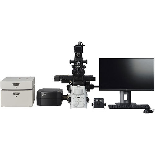
Tell us about your requirement

Price:
Quantity
Select Unit
- 50
- 100
- 200
- 250
- 500
- 1000+
Additional detail
Mobile number
Email

More Products in Nanotech Lab Category
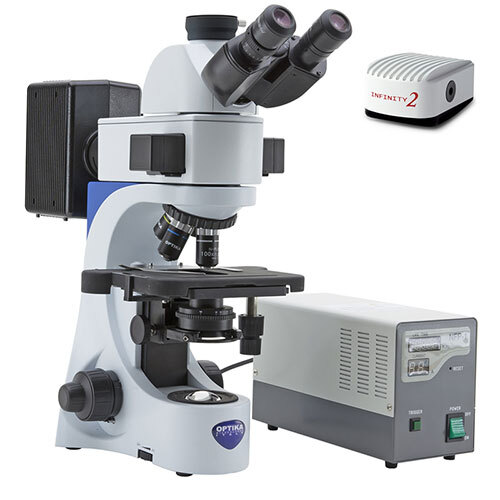
Fluorescence Microscope
Minimum Order Quantity : 1 Piece
Warranty : 1 Year
Usage : Laboratory
Material : Metal & Plastic
Type : Other, Fluorescence Microscope
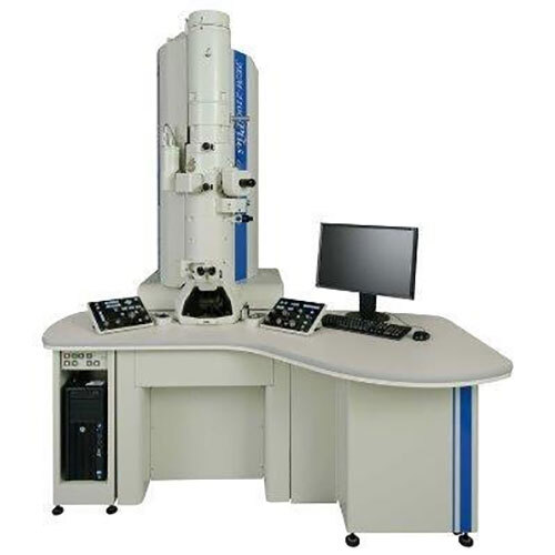
TRANSMISSION ELECTRON MICROSCOPE
Minimum Order Quantity : 1 Piece
Warranty : 1 Year
Usage : Laboratory
Material : Mild Steel
Type : Other, TRANSMISSION ELECTRON MICROSCOPE
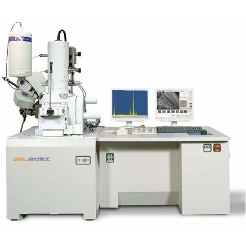
SCANNING ELECTRON MICROSCOPE
Minimum Order Quantity : 1 Piece
Warranty : 1 Year
Usage : Laboratory
Material : Mild Steel
Type : Other, SCANNING ELECTRON MICROSCOPE
Our Products
- Quality Control Lab
- Biotechnology Lab
- Life Science & Biochemistry
- Nanotech Lab
- Medical & Biomedical Lab
- Metal Analyzing
- Pharmacy Lab
- Industrial Chemicals
- Foundry Alloys & Metals
- Micronutrients For All Crops
- Glasswares
- Lab Furniture
- EDP Equipments
- LABORATORY CHEMICALS
- Mechanical engineering
- Civil engineering
- Electrical/Electronics engineering
- Electronics & Communication engineerings
- Textile Technology
- aerospace/aeronautical engineering
- Automobile engineering

160, 6th Street Extension, 100 feet road, Gandhipuram, Coimbatore - 641012, Tamil Nadu, India

Mr C. S. R. Cholan
(Chief Executive Officer)
Mobile :07971405282
 Send Inquiry
 Send SMS
Developed and Managed by Infocom Network Private Limited.
